
Jerry Fuh
Jerry Fuh, PhD
Professor
the Department of Mechanical Engineering, National University of Singapore
Email: jerry.fuh@nus.edu.sg
Biography:
Dr. Jerry Fuh is a Professor at the Department of Mechanical Engineering, National University of Singapore (NUS) and the Co-Director of NUS Centre for Additive Manufacturing (AM.NUS) focused on biomedical technologies. He is a Fellow of SME and ASME and a PE from California, USA. Dr. Fuh has devoted himself to the research of Additive Manufacturing (AM) processes or 3D Printing (3DP) since 1995. He and his colleagues have established the NUS’s AM/3DP research programme focusing on biomedical applications and set up an advanced 3DP laboratories through several research & development grants with industrial collaborations.
In 2005, he received the IES Prestigious Engineering Achievement Award for the work on “Development of Rapid Prototyping Technologies for Precision and Biomedical Engineering” from the Institute of Engineers, Singapore (IES) in recognition of outstanding engineering skills which have made notable contributions to progress engineering in Singapore. He has published over 350 technical papers in advanced manufacturing and design, and supervised over 80 graduate students with over 50 are PhD students. He also serves in more than 10 refereed journals as Editor, Associate Editor or Editorial Board Members related to design, manufacturing and AM/3DP
Abstract:
E-Jet Bioprinting of Hierarchical Scaffold for Esophagus Tissue Repair
Esophagus is a muscular tuber in which food and drink are transported from pharynx to stomach. However, the function of esophagus could be damaged due to diseases. To rebuild the damaged esophagus, a suitable replacement is needed to re-establish the function of esophagus. The muscular wall of esophagus was found in bilaminar arrangement: an inner layer where fibroblasts oriented circumferentially to lumen axis and an outer layer where cells aligned parallel to lumen axis. This arrange-ment is essential in propelling food boluses or fluid. In detail, the sequential contrac-tion of the circular muscle layer occludes the lumen to push the ingested bolus longi-tudinally along the esophagus, and the sequential contraction of the longitudinal mus-cle shortens the esophagus tract. The contraction of longitudinal muscle layer also in-creases the density of the circular muscle fibers and, in turn, increases the efficiency of the circular muscle contraction.
Recently, artificial biopolymers such as Poly(L-lactide-co-caprolactone) (PLLC) and polycaprolactone (PCL) were applied to fabricate esophageal tissue engineering scaffolds. However, due to the intrinsic defects of fabrication, the structures of previ-ous scaffolds are not regular. Hence, they can hardly orient cells regular alignment, which provides poor replicate to the natural esophageal structure.
Porous scaffolds could facilitate cells connections as well as nutrition exchange, hence, promoting cells’ growth and proliferation. However, the pores also influence cell seeding efficiency. When cells are pipetted on top of scaffolds, many cells could go through the pores directly and left on the bottom of culture plate. Subsequently, it takes a long time for these cells to proliferate and adhere onto the fibers of scaffold, which leads to a waste of cells seeding and culturing time.
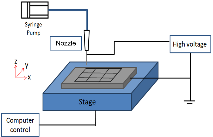
The objective of this study is to fabricate hierarchical esophageal tissue engineer-ing scaffold to guide cellular regular orientation as well as to increase cell adhesion, growth and proliferation hence, mimicking the structure of natural esophagus. Electro-hydrodynamic jetting (E-jetting) and electro-spraying (e-spraying) were adopted to fabricate the scaffold (Figure 1). Furthermore, human esophagus fibroblasts (HEF) were cultured on this scaffold to determine the function of fibre cell alignment.
The effects of different scaffolds to influence cellular orientation was demon-strated through cytoskeletal staining tests. Figure 4(a) shows the F-actin and nucleus morphology of HEsF on different scaffolds. The orientations of HEsF on the e-jetted scaffold were mainly oriented along the longitudinal fibers. On the other hand, the HEsF on hierarchical scaffolds and e-sprayed film orientated in random directions. The HEsFs with nuclei angles between 0 - 20 ° and 160 - 180 ° were designated as aligned cells. Figure 4(b) shows the nuclei angles distribution on each group. On e-jetted scaffolds, the fibroblasts nuclei angles were mainly between the range of 0-20 ° and 160 °-180 °. On the hierarchical scaffold, however the nuclei angle distributed more evenly. Meanwhile, the nuclei angle was focused more on the range of 60 °-80 °.

Professor
the Department of Mechanical Engineering, National University of Singapore
Email: jerry.fuh@nus.edu.sg
Biography:
Dr. Jerry Fuh is a Professor at the Department of Mechanical Engineering, National University of Singapore (NUS) and the Co-Director of NUS Centre for Additive Manufacturing (AM.NUS) focused on biomedical technologies. He is a Fellow of SME and ASME and a PE from California, USA. Dr. Fuh has devoted himself to the research of Additive Manufacturing (AM) processes or 3D Printing (3DP) since 1995. He and his colleagues have established the NUS’s AM/3DP research programme focusing on biomedical applications and set up an advanced 3DP laboratories through several research & development grants with industrial collaborations.
In 2005, he received the IES Prestigious Engineering Achievement Award for the work on “Development of Rapid Prototyping Technologies for Precision and Biomedical Engineering” from the Institute of Engineers, Singapore (IES) in recognition of outstanding engineering skills which have made notable contributions to progress engineering in Singapore. He has published over 350 technical papers in advanced manufacturing and design, and supervised over 80 graduate students with over 50 are PhD students. He also serves in more than 10 refereed journals as Editor, Associate Editor or Editorial Board Members related to design, manufacturing and AM/3DP
Abstract:
E-Jet Bioprinting of Hierarchical Scaffold for Esophagus Tissue Repair
Esophagus is a muscular tuber in which food and drink are transported from pharynx to stomach. However, the function of esophagus could be damaged due to diseases. To rebuild the damaged esophagus, a suitable replacement is needed to re-establish the function of esophagus. The muscular wall of esophagus was found in bilaminar arrangement: an inner layer where fibroblasts oriented circumferentially to lumen axis and an outer layer where cells aligned parallel to lumen axis. This arrange-ment is essential in propelling food boluses or fluid. In detail, the sequential contrac-tion of the circular muscle layer occludes the lumen to push the ingested bolus longi-tudinally along the esophagus, and the sequential contraction of the longitudinal mus-cle shortens the esophagus tract. The contraction of longitudinal muscle layer also in-creases the density of the circular muscle fibers and, in turn, increases the efficiency of the circular muscle contraction.
Recently, artificial biopolymers such as Poly(L-lactide-co-caprolactone) (PLLC) and polycaprolactone (PCL) were applied to fabricate esophageal tissue engineering scaffolds. However, due to the intrinsic defects of fabrication, the structures of previ-ous scaffolds are not regular. Hence, they can hardly orient cells regular alignment, which provides poor replicate to the natural esophageal structure.
Porous scaffolds could facilitate cells connections as well as nutrition exchange, hence, promoting cells’ growth and proliferation. However, the pores also influence cell seeding efficiency. When cells are pipetted on top of scaffolds, many cells could go through the pores directly and left on the bottom of culture plate. Subsequently, it takes a long time for these cells to proliferate and adhere onto the fibers of scaffold, which leads to a waste of cells seeding and culturing time.
The objective of this study is to fabricate hierarchical esophageal tissue engineer-ing scaffold to guide cellular regular orientation as well as to increase cell adhesion, growth and proliferation hence, mimicking the structure of natural esophagus. Electro-hydrodynamic jetting (E-jetting) and electro-spraying (e-spraying) were adopted to fabricate the scaffold (Figure 1). Furthermore, human esophagus fibroblasts (HEF) were cultured on this scaffold to determine the function of fibre cell alignment.
Figure 1. Schematic of E-jetting and E-spraying process
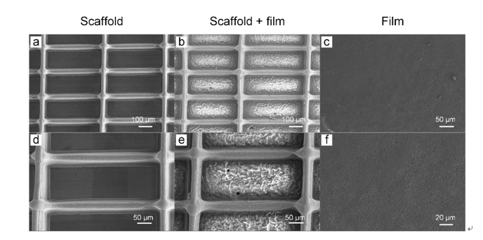
Figure 2. SEM images of (a, d) e-jetted scaffold, (b, e) hierarchical scaffold and
(c, f) e-sprayed film.
Primary human esophagus fibroblasts (HEsF #2730) were seeded on the scaffolds (Figure 2). There are only few dead cells on all three kinds of scaffolds in all time points, which proves the good biocompatibility of all scaffolds. Furthermore, with the increase of culturing time, HEsF spread and proliferated on the surface of scaffolds gradually. On the hierarchical scaffold, fibroblasts covered the inter-fiber area, while there were some round HEsF cells on the film (Figure 3).
The results show that hierarchical scaffold enhanced cell alignment and adhesion, indicating its potential in the applications to esophageal tissue and other tissues with similar struc-tures such as tendon.
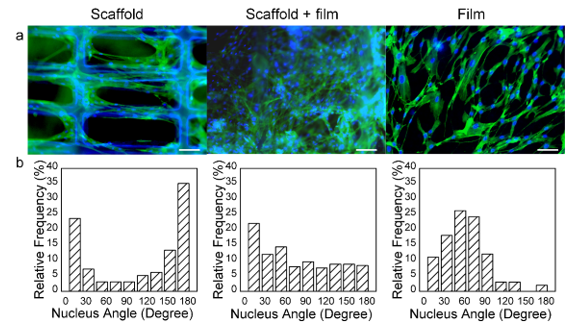
The effects of different scaffolds to influence cellular orientation was demon-strated through cytoskeletal staining tests. Figure 4(a) shows the F-actin and nucleus morphology of HEsF on different scaffolds. The orientations of HEsF on the e-jetted scaffold were mainly oriented along the longitudinal fibers. On the other hand, the HEsF on hierarchical scaffolds and e-sprayed film orientated in random directions. The HEsFs with nuclei angles between 0 - 20 ° and 160 - 180 ° were designated as aligned cells. Figure 4(b) shows the nuclei angles distribution on each group. On e-jetted scaffolds, the fibroblasts nuclei angles were mainly between the range of 0-20 ° and 160 °-180 °. On the hierarchical scaffold, however the nuclei angle distributed more evenly. Meanwhile, the nuclei angle was focused more on the range of 60 °-80 °.
Figure 3. Viability of HEsF on esophageal tissue engineering scaffolds after culturing for 1, 3 and 7 days. Live cell: green color, dead cells: red color. Scale bar: 100 μm
Figure 4. (a) Cytoskeletal staining of HEsF on three kinds of scaffolds after 7 days. Phalloidin: green color, F-actin; DAPI: blue color, nucleus. (b) Nucleus angle
distribution.